
3D labels your lab can reuse
Connect 3D labelling and review to the exact dataset your paper uses, so collaborators can inspect the work behind each segmentation.
Photograph: Peter Smyth, Wikimedia Commons, CC0 (opens in a new tab), graded and small stickers blurred. Sources
Why it matters
The paper needs more than a mask folder.
Keep the source scan, label revisions and class definitions together, so another researcher can inspect what your analysis used.
- A navel orange, rendered from its food X-ray CT scan.
- A plane opens the scan at the equator, slice 358.
- Slice 358, in the scan's own greys.
- Short dashes: SAM 2.1's proposal, from a recorded run outside Nitsor.
- Long dashes: the authors' scripted pulp segmentation, shipped with the scans. No person drew it.
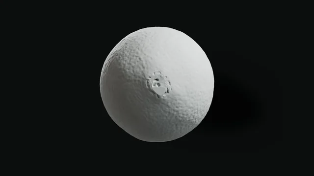
1/5A navel orange, rendered from its food X-ray CT scan.
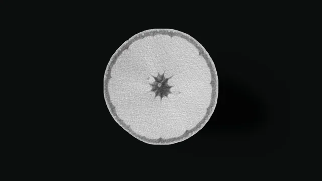
2/5A plane opens the scan at the equator, slice 358.
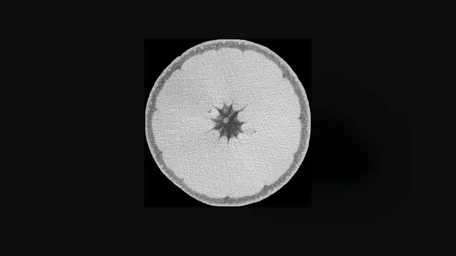
3/5Slice 358, in the scan's own greys.
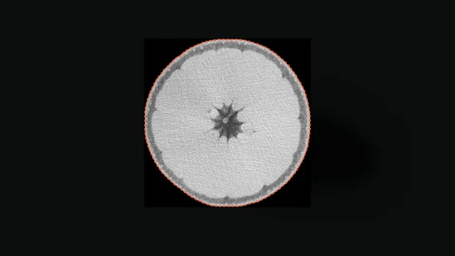
4/5Short dashes: SAM 2.1's proposal, from a recorded run outside Nitsor.
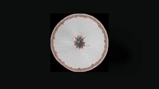
5/5Long dashes: the authors' scripted pulp segmentation, shipped with the scans. No person drew it.


Real X-ray CT of a navel orange: Amézquita et al., Michigan State University, CC0 (opens in a new tab), modified (rendered, cut, windowed, masks drawn dashed). Long dashes: the authors' scripted segmentation (thresholding and morphology, checked by eye), shipped with the scans; no person drew it. Short dashes: model proposal from SAM 2.1, Meta AI, Apache 2.0 (opens in a new tab), run outside Nitsor.
How it works here
Give your methods a fixed dataset.
A lab group can register a volume, label it, have a second person review the work, and fix the dataset version the paper cites.
Register
Register the reconstruction.
Micro-CT, cone-beam CT and MRI volumes register from your S3-compatible storage, with spacing kept per axis.
Label
Label in 3D.
The same brush, contour and threshold tools, at the scale the instrument recorded.
Review
A second person reviews.
They accept, correct or reject each label, and the log keeps both names.
Pin
Pin a dataset version.
It fixes the labels your paper used.
What the record holds
What another lab needs to reuse your labels.
Read-only access, the dataset version, and the history of every label in it, in the workspace or through the API.
Cited in the methods
- Version
- dsv_2c8e51f0
- Volumes
- 3 micro-CT scans
- Drawn by
- H. Abara, annotator
- Reviewed by
- T. Lindgren, second reader
- Bound to
- 0d9a61fe
Worked example on public scans. Names, decisions and version IDs are examples.
Spacing kept on every axis
Nitsor reads spacing, orientation and origin from the file header and keeps each series on its own grid.
Review the exact label revision
Each label names its author. A review records who checked it and which revision they saw.
Keep published inputs fixed
A dataset version is frozen, with a digest for every chunk, so a read can be checked against it. Existing scans stay in their connected storage; uploads use your chosen destination. The browser reads viewed slices through short-lived signed links, and model jobs use temporary copies.
What opens
Reconstructed volumes as DICOM Part 10 (uncompressed) or VGStudio .vgi/.vol, from the tools you already use.
What gets scanned
Wood and plant anatomy
Cell walls, vessels and fibres.
Seeds, grain and nuts
On MRI and X-ray CT.
Rock cores
Pores and grains in a core.
Foams and fibre composites
Cells, struts and fibres.
Battery materials
Electrodes and their layers.
Worked example
An orange and an inner ear, as their authors segmented them.
A navel orange on X-ray CT, with rind, pulp and oil glands segmented by the lab that scanned it, with thresholding and morphology scripts, and a human inner ear on micro-CT, with an expert segmentation and five landmarks placed by an otologist.

Navel orange, food X-ray CT: the authors' scripted segmentation
- Tissues
- rind, pulp, central column
- Oil glands on this slice
- 96, derived
- Oil glands in the fruit
- 8,358, derived
Dashed: the authors' scripted segmentation (thresholding and morphology, checked by eye), shipped with the scans; no person drew it. Gland circles and counts are derived from it.
Real X-ray CT: Amézquita et al., Michigan State University, CC0 (opens in a new tab), modified (one slice windowed and cropped, masks drawn dashed, glands counted).

Human bony labyrinth, micro-CT: expert segmentation and five landmarks
- RW
- centre of the round window
- V
- centre of the vestibule
- OW
- centre of the oval window
- C
- centre of the cochlear basal turn
- A
- helicotrema
An expert labelled the segmentation by hand, with region growing, thresholding and manual correction, and an experienced otologist reviewed it. An otologist placed the landmarks.
Real micro-CT: Wimmer et al., Bern University Hospital and Inria, CC BY 4.0 (opens in a new tab), modified (surface rendered from the published label, landmarks projected).
Questions
Before you choose Nitsor
- Our slices are further apart than our pixels. Does that survive?
- Yes. Nitsor reads spacing, orientation and origin from supported file headers, including different spacing along each axis.
- Can another lab check our labels?
- Invite them read-only, with no per-person charge. They read the dataset version, its labels and their history in the workspace or through the API.
- Can we script it?
- Yes. The HTTP API covers registration and dataset versions, and you call it with a scoped agent credential issued in the hosted app.
- Does Nitsor reconstruct projections?
- No. Bring the reconstructed volume from your reconstruction software.
Working with uncompressed single-frame DICOM ultrasound images?
Label and review uncompressed single-frame DICOM ultrasound images. Calibration stays with the image; images without spatial geometry remain individual images.
Explore ultrasound researchStart with one volume.
See how a public micro-CT, cone-beam CT or MRI volume becomes a labelled, reviewed dataset you can cite.