
Real footage · NASA materials evaluation lab
Real footage · NASA materials evaluation lab
Train inspection AI
on reviewed 3D scans.
Label CT and MRI scans, review model suggestions and keep the exact dataset behind each training run.
Public scans, model proposals and licensed footage. Sources
Scroll to exploreOne scan, layer by layer.
A real scan, what a model computed and proposed from it, the label people published, and the record that keeps them apart.
Aerospace · industrial CTFraunhofer sample V5, a historic airframe section, axial slice 300
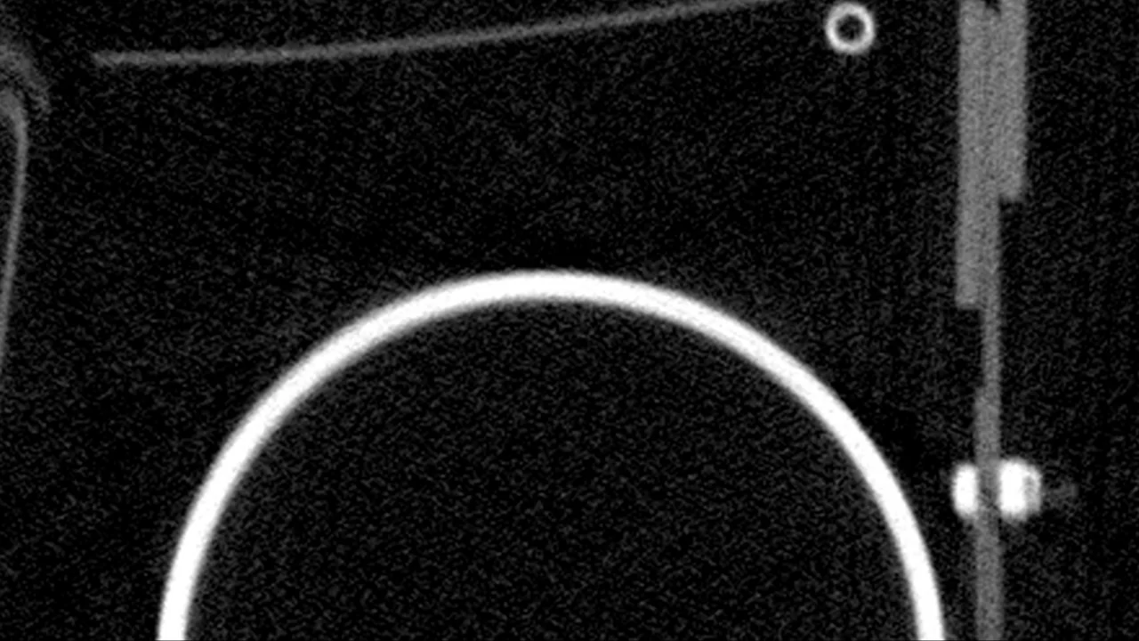

01 Scan slice
Axial slice 300 of the real volume, windowed: the model's input, with the one box it was given.
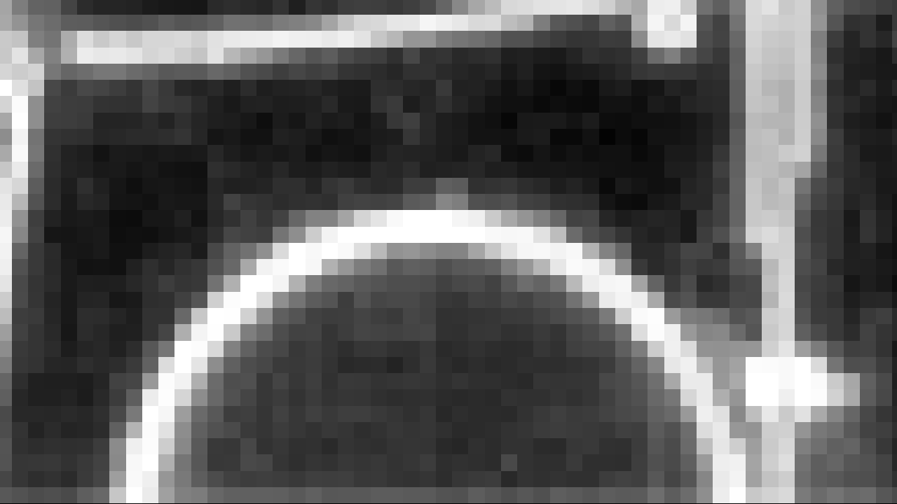
02 Encoder features, PC1
SAM 2.1's image encoder turns the slice into 256 channels on a 64 x 64 grid. This is their first principal component (17% of the variance), at its native 16 px blocks. A projection, not a layer.
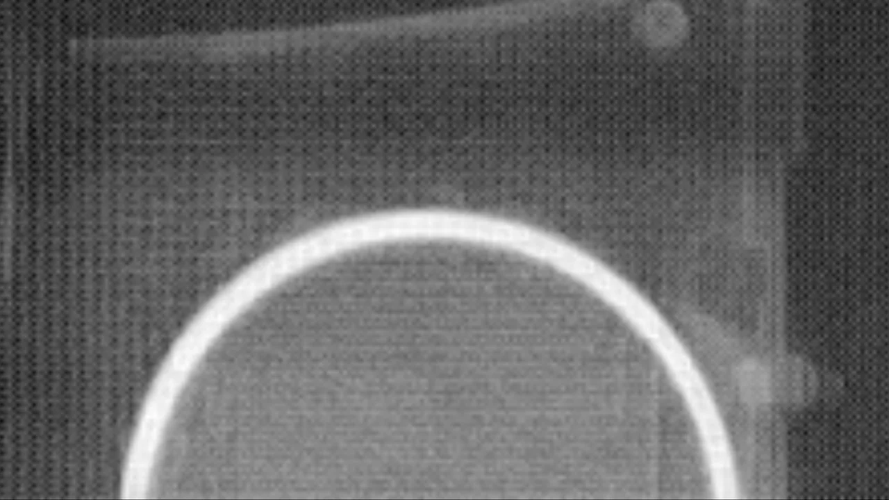
03 Mask logits
The mask decoder's output, -25 to +8. Logits are not probabilities.
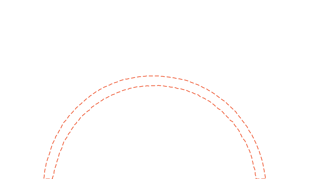
04 Model proposal
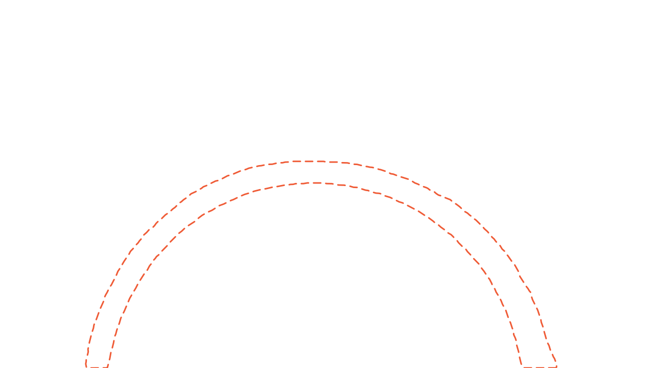
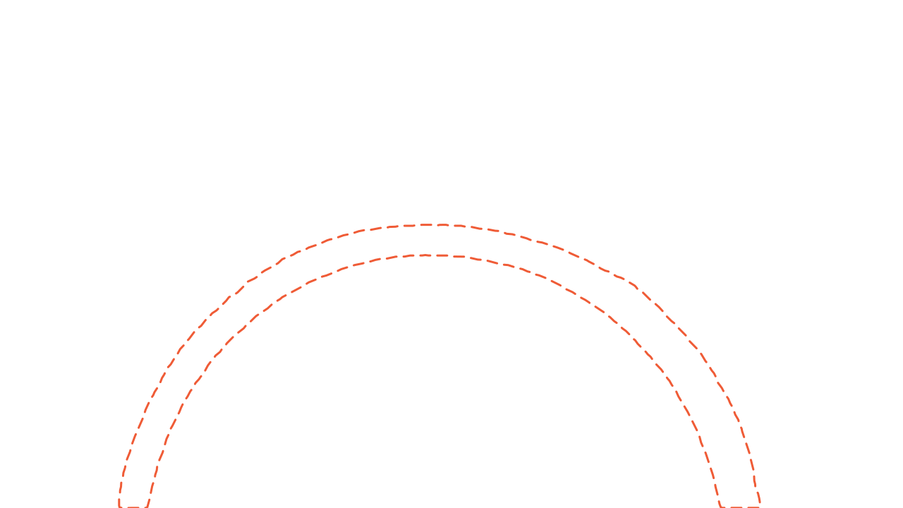
Where the logits are above zero: SAM 2.1's outline of the bottle wall, dashed.

05 Published label
Instance 138, the pressure bottle, as published by the dataset's annotators (solid), with neighbouring parts' published instance outlines (thin). IoU with the proposal: 0.82.
Labels on the picture: 148 · 146 · 140 · 139- Aerospace · V5scan fba9a5cfSAM 2.1 proposal 68d34799, on its own branchpublished label 138 1c7047f3, merged
- dataset main, tag dv-sample-01, read by the training input preview
06 Record
The history for this scan: the proposal stays on its own branch, each published human label joins the dataset line on its own, and a tag pins the version a training job reads.


CT and instance masks: Gruber et al., Fraunhofer EZRT, doi 10.5281/zenodo.10651746, CC BY 4.0 (opens in a new tab). Model: SAM 2.1 hiera-tiny, run by Nitsor outside the product. Modified by Nitsor: windowed, cropped, outlines and model outputs drawn over it.
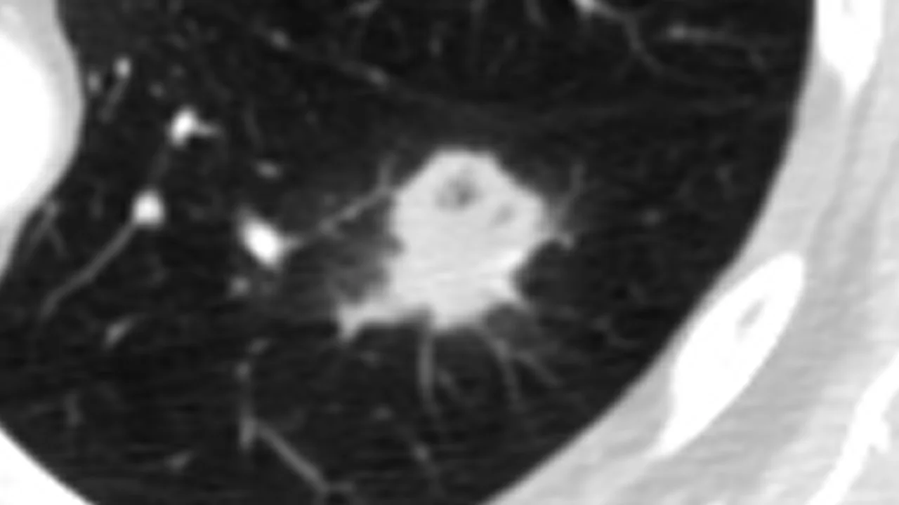
Healthcare · clinical CTLIDC-IDRI-0003, axial slice 64, nodule field about 112 x 63 mm
01 Scan slice
Slice 64 of a public chest CT, lung window. The same patient the four radiologists outlined. The box given to the model is the readers' combined extent plus 3 mm, derived from their outlines.

02 Encoder features, PC1
SAM 2.1's image encoder, first principal component of 256 channels (25% of the variance), native 16 px blocks.
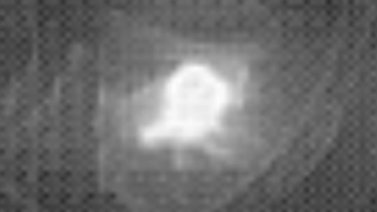
03 Mask logits
SAM 2.1's decoder output for one box around the nodule. Logits, not probabilities.

04 Lung lobes, a second model
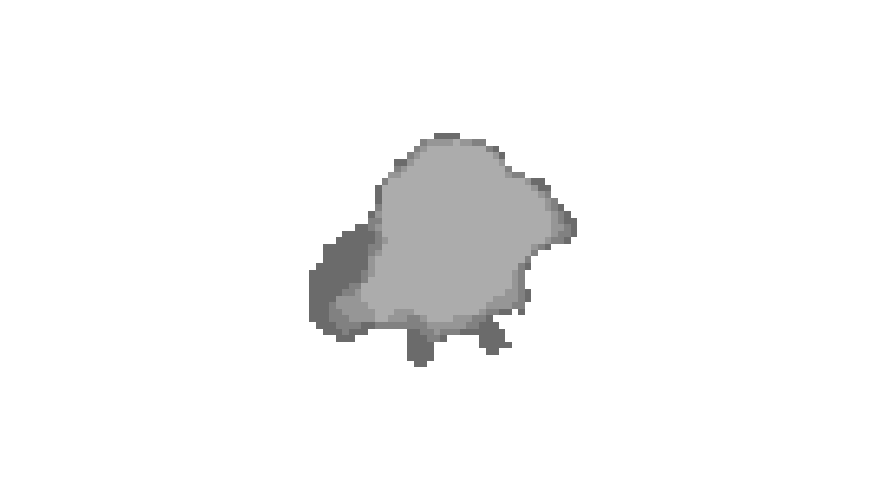
A second model, a different task: lung lobes. TotalSegmentator's softmax for the class 'left lower lobe' on the same field, per voxel. It does not look for the nodule.
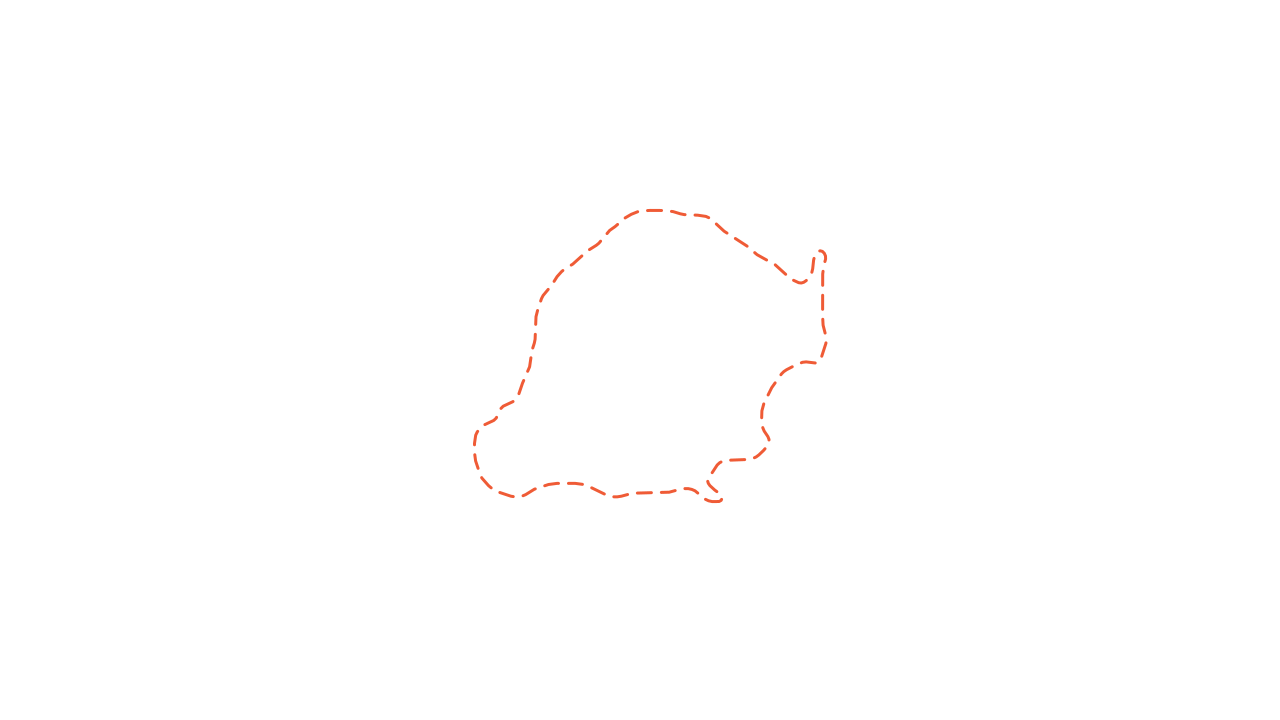
05 Model proposal
SAM 2.1's outline of the nodule, dashed. IoU with the four readers: 0.79, 0.80, 0.84, 0.84.





06 Four published outlines
Four radiologists' outlines, kept separate, over their agreement (grey, 1 to 4). Longest diameter of the 2-of-4 consensus: 32.8 mm, derived from the outlines.
- Healthcare · LIDC-0003scan 58628ab1SAM 2.1 proposal 96cab4b8, on its own branchreader 1 aa3e882e, mergedreader 2 9e9a9479, mergedreader 3 55e99aaa, mergedreader 4 1a30ac07, merged
- dataset main, tag dv-sample-01, read by the training input preview
07 Record
The history for this scan: the proposal stays on its own branch, each published human label joins the dataset line on its own, and a tag pins the version a training job reads.


32.8 mm, longest diameter of the 2-of-4 consensus, derived from the published outlines
CT and reader outlines: Armato et al., "Data From LIDC-IDRI", TCIA, doi 10.7937/K9/TCIA.2015.LO9QL9SX, CC BY 3.0 (opens in a new tab). Models: SAM 2.1 hiera-tiny and TotalSegmentator v2, run by Nitsor outside the product. Modified by Nitsor: windowed, cropped, outlines and model outputs drawn over it.
Healthcare · MRISPIDER case 101, sagittal T2 MRI, slice 8
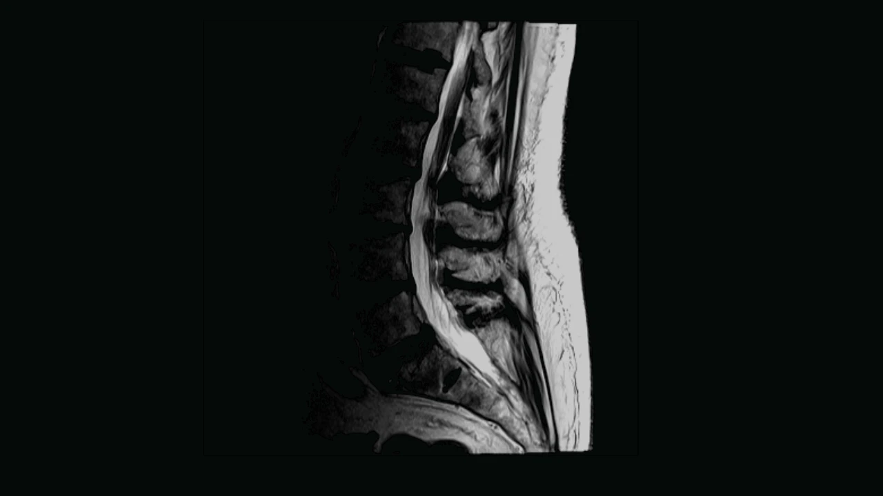
01 MRI slice
A sagittal T2 image from SPIDER case 101, slice 8 across. The display keeps the scan's own voxel spacing; the image and the masks share one geometry.
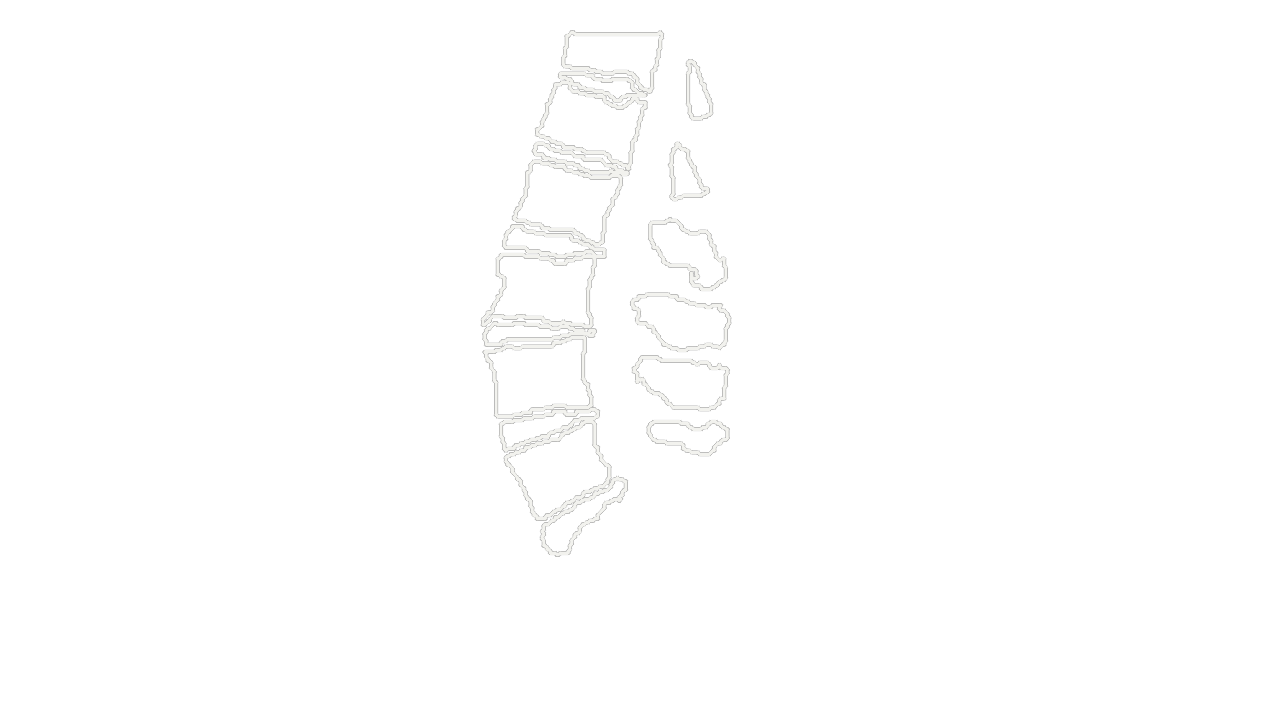
02 Published masks
Solid white: the published vertebra (01 to 06) and disc (201 to 206) masks on this slice. The dataset made them semi-automatically, then its annotators reviewed and manually corrected them. The numbers count from the bottom up; they are not anatomical names.
Lumbar MRI and reference masks: SPIDER, van der Graaf et al., Radboud University Medical Center, doi 10.5281/zenodo.10159290, CC BY 4.0 (opens in a new tab). Modified by Nitsor: cropped, and the published masks drawn as outlines. No model output is drawn on this scan.
Food · fruit CTNavel orange SW03, CT slice 358, the equator
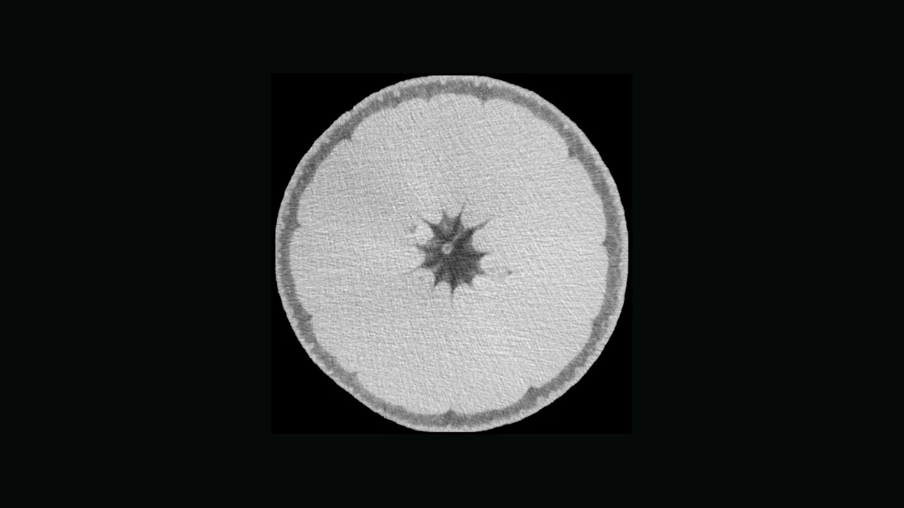
01 Scan slice
The equatorial slice of a real orange CT: rind, pulp segments and the central column.
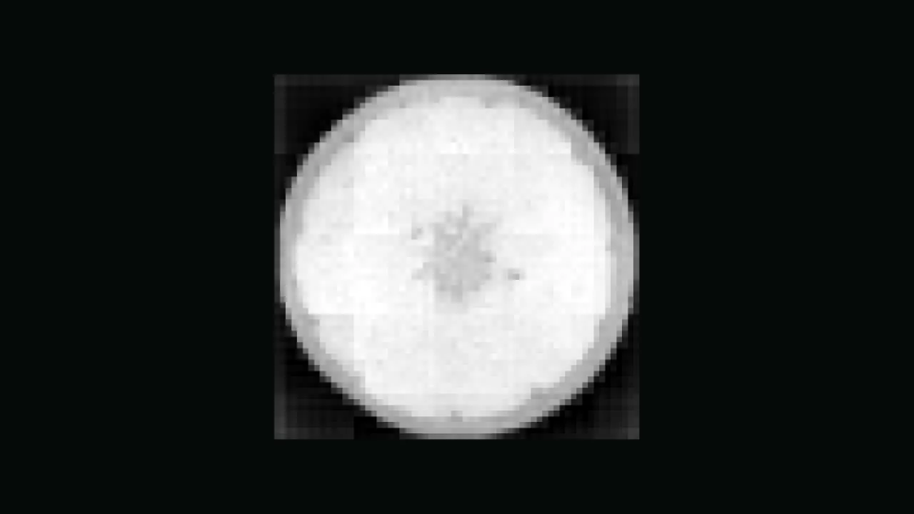
02 Encoder features, PC1
SAM 2.1's image encoder, first principal component of 256 channels (32% of the variance), at its native blocks.
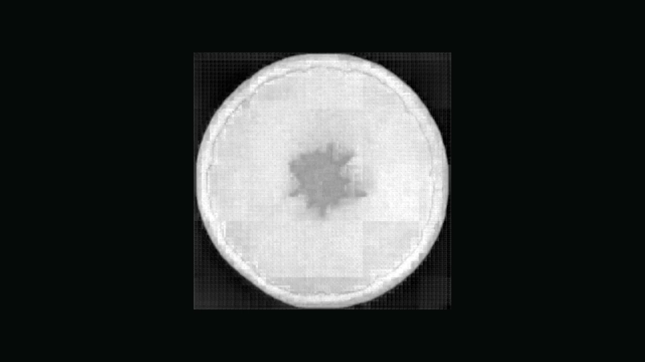
03 Mask logits
The decoder's output for one box, -9 to +7. Logits are not probabilities.
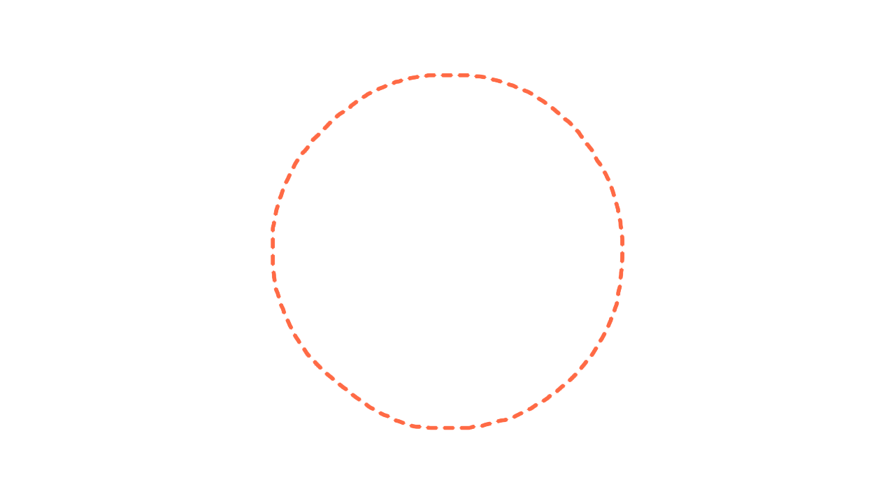
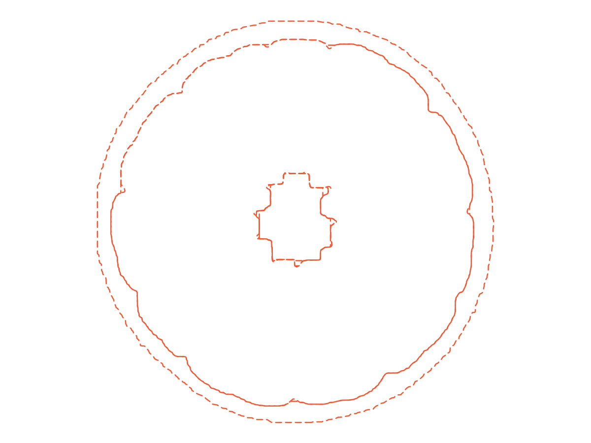
04 Model proposal
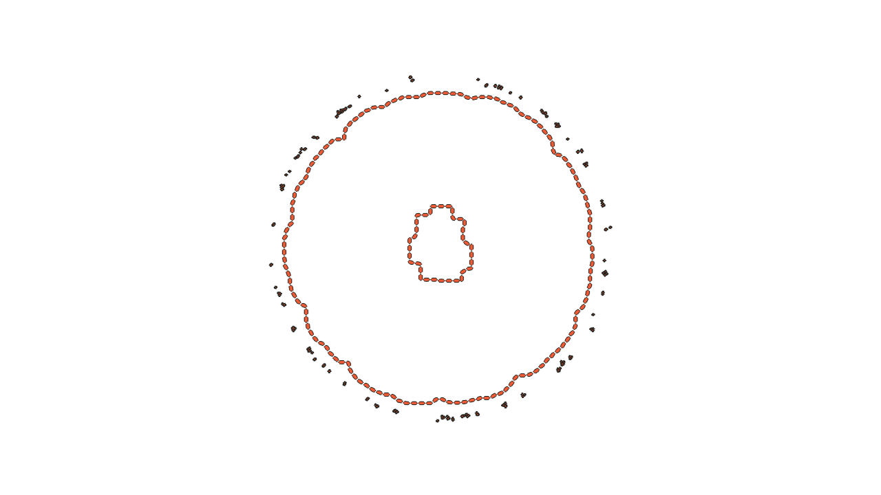
SAM 2.1's recorded proposal, long dashes. It takes in the rind; the authors' scripted pulp mask stops inside it.
05 Authors' scripted masks
Short dashes: the pulp and oil-gland masks, the authors' scripted segmentation (thresholding and morphology, parameters set by hand for each fruit and checked by eye), shipped with the scans; no person drew them. IoU 0.76 compares SAM 2.1 with the scripted pulp mask, not with a human label.
Citrus CT and its scripted masks: Amézquita, Quigley, Ophelders, Seymour, Munch, Chitwood, doi 10.5061/dryad.34tmpg4n6, CC0 (opens in a new tab). Model: SAM 2.1 hiera-tiny, run by Nitsor outside the product. Modified by Nitsor: cropped, and SAM 2.1's proposal and the authors' scripted masks drawn as dashed outlines.
Hashes: sha256 prefixes of the published files.
See the workspace

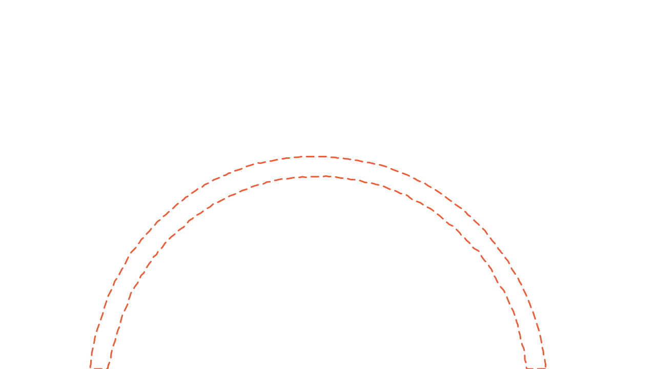
Slice 300, one box

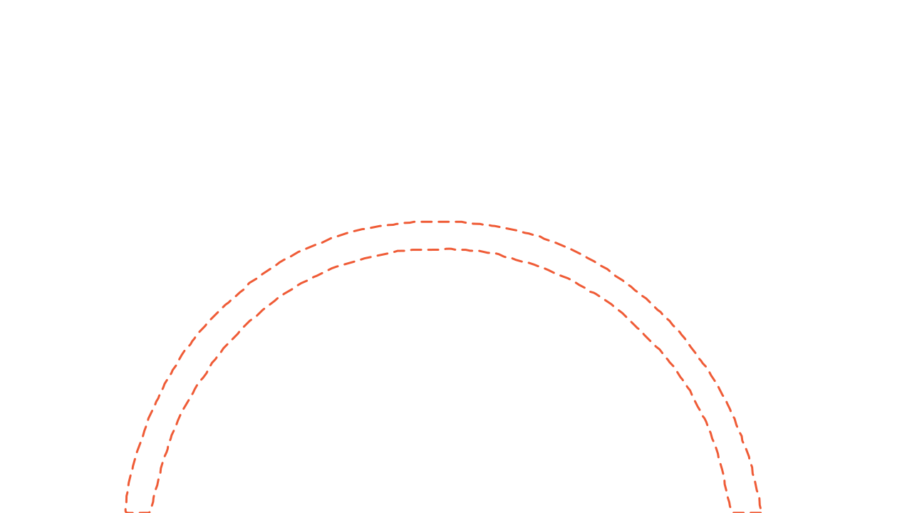
Slice 316

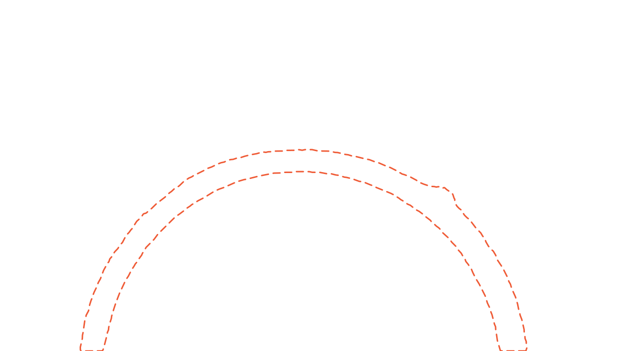
Slice 332

Slice 348

Slice 360
Proposal
- Proposed by
- SAM 2.1, from one box
- Slices
- 64, from slice 300
- State
- recorded, awaiting review
Model proposals from SAM 2.1 (Apache 2.0 (opens in a new tab)), run outside Nitsor. Real industrial CT, Gruber et al., Fraunhofer EZRT, CC BY 4.0 (opens in a new tab), modified: windowed, cropped, the proposals drawn over it.
Proposals
Review model suggestions against the scan.
Models and agents are first-class in Nitsor. Mark one slice and a model proposes masks on up to 64 slices; reviewers accept, correct or reject each one instead of drawing it.
See model proposals

Review, one nodule
- Proposed by
- SAM 2.1, from one box
- Outlined by
- four radiologists, kept separate
- Longest diameter
- 32.8 mm, derived
- Reviewer
- P. Kowalski
The outlines are the LIDC readers' own, as published.
Four published reader outlines and a model proposal from SAM 2.1 (Apache 2.0 (opens in a new tab)), outside Nitsor. The box the model was given is the readers' combined extent plus 3 mm, derived from their outlines. Real chest CT, Armato et al., "Data From LIDC-IDRI", TCIA, CC BY 3.0 (opens in a new tab), modified: lung-windowed, cropped, the outlines redrawn, the agreement map and diameter derived.
Review
See which revision the reviewer checked.
A review stays attached to the label revision inspected. Rejected work stays visible, nobody reviews their own, and uncertainty scores can send the least certain work to reviewers first.
See how review works- Aerospace · V5scan fba9a5cfSAM 2.1 proposal 68d34799, on its own branchpublished label 138 1c7047f3, merged
- Healthcare · LIDC-0003scan 58628ab1SAM 2.1 proposal 96cab4b8, on its own branchreader 1 aa3e882e, mergedreader 2 9e9a9479, mergedreader 3 55e99aaa, mergedreader 4 1a30ac07, merged
- Food · apple 98scan cb1c419aSAM 2.1 proposal 14bc3a32, on its own branchbrowning score 10/10, no mask f1c1903d, joined as a score, with no mask
- dataset main, tag dv-sample-01, read by the training input preview
Real scans and published labels, modified (windowed, cropped, outlines drawn): Gruber et al., Fraunhofer EZRT, CC BY 4.0 (opens in a new tab); Armato et al., "Data From LIDC-IDRI", TCIA, CC BY 3.0 (opens in a new tab); Schut et al., CWI and GREEFA, CC BY 4.0 (opens in a new tab). Proposals from SAM 2.1 (Apache 2.0 (opens in a new tab)), outside Nitsor. Hashes: sha256 prefixes of the published files.
Datasets
Keep training data fixed as labels change.
Give each training job a fixed dataset version. It keeps reading the same scans and labels while your team continues editing.
See versioningThe system of evidence for AI-assisted inspection
Identify the scans, labels and reviews behind each training dataset.

What each frame shows
- Step through the slices of a chest CT
- Select a label to find it on the scan
- Each label is saved with its scan and slices
Real chest CT: Armato et al., "Data From LIDC-IDRI", TCIA, CC BY 3.0 (opens in a new tab), modified: cropped. Labels from the LIDC readers' published annotations.


Agents call the API with a credential of their own.
Real open scans. Sources
Why Nitsor
When someone asks which labels trained the model, you have an answer.
Who changed this label, and who checked it?
Every revision keeps its author, and each verdict on a label records the version of the label it judged.
Where do our scans go?
In your storage. The browser reads viewed slices from there through short-lived signed links, and model jobs use temporary copies.
Do we pay for every reviewer?
No. No plan charges per person, on Community, Team or Enterprise.
Solutions
For teams working with CT and MRI.
Castings, welds, composites, material samples and research specimens, with every label and review tied to the scan it was drawn on.
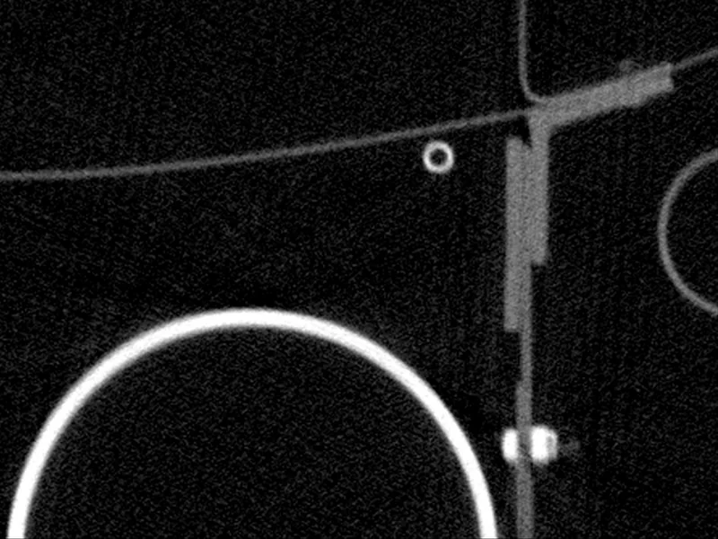
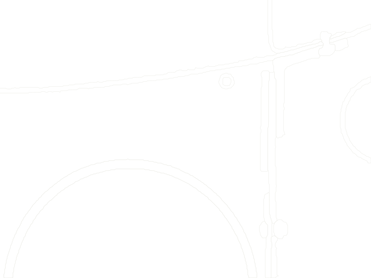 Aerospace · industrial CTInstance segmentation: every part keeps its own published ID.12 published instances in view, slice 300Labels on the picture: 17 · 19 · 23 · 35 · 78 · 121 · 138 · 139 · 146 · 148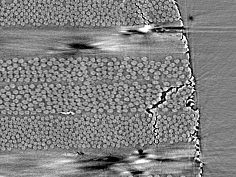
 Composites · synchrotron CTDefect segmentation: three classes of ply damage, traced by people.Damage traced by trained annotators, published with the scans · specimen 2, load step eLabels on the picture: 0° ply damage · ±45° ply damage · 90° ply damage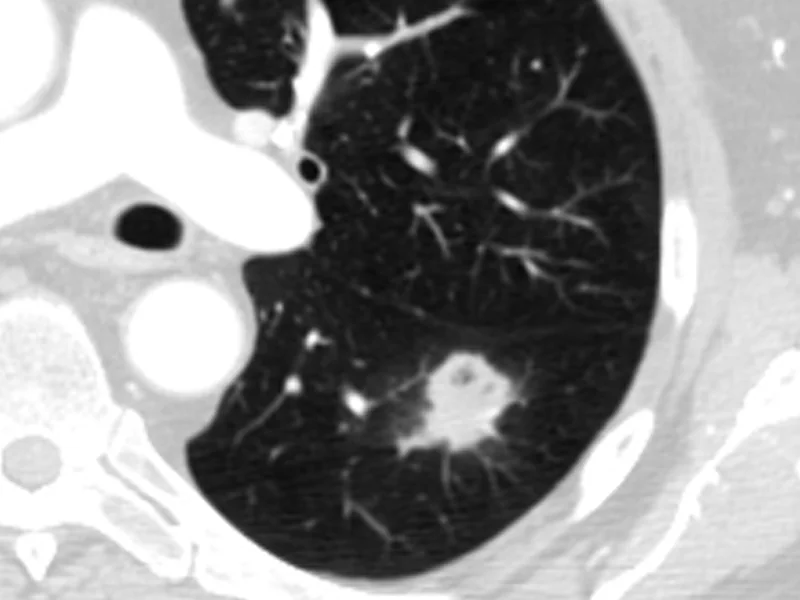

 Healthcare · clinical CTFour radiologists' outlines, and the 3D box they imply.3D box 30.4 x 26.2 x 17.5 mm, derived from the published outlines (at least 2 of 4 readers, 7 slices)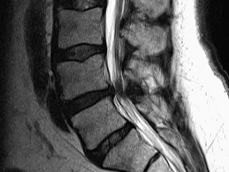
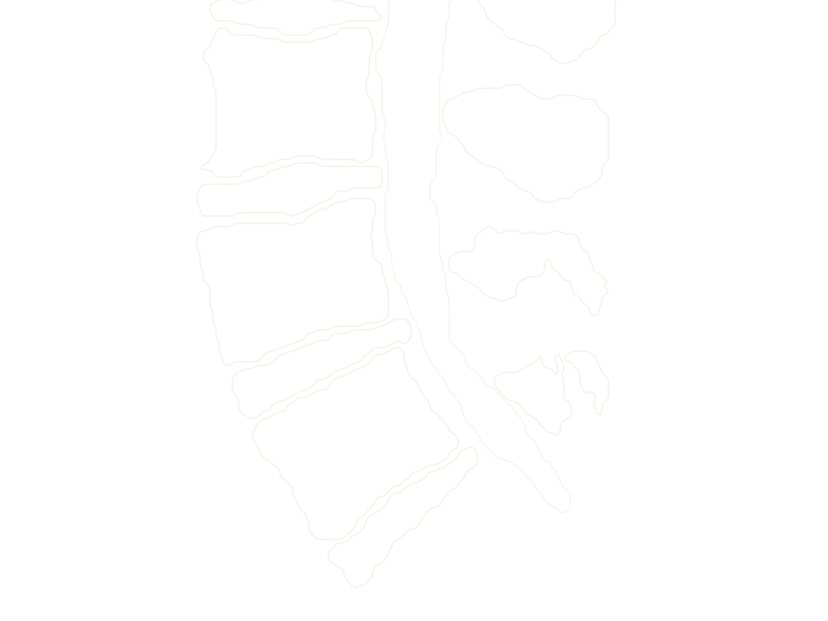 Healthcare · MRIVertebra, disc and canal masks, and a grade for every disc.P: Pfirrmann grade 1 to 5, scored per disc by a musculoskeletal radiologist (published)Labels on the picture: P4 · P4 · P3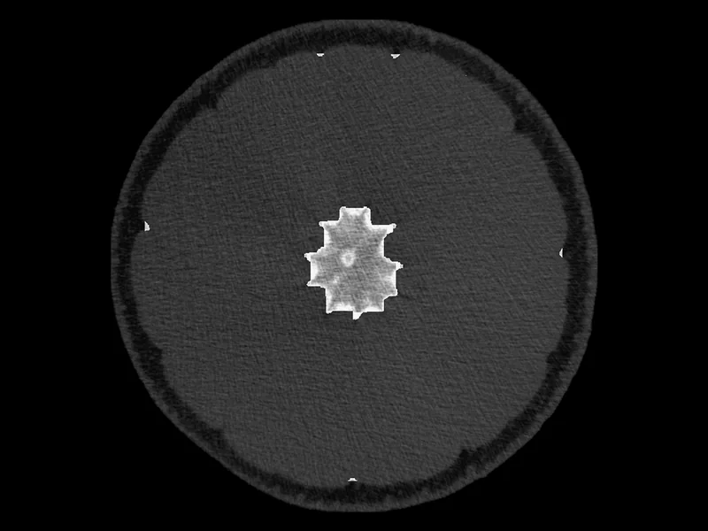
 Food · X-ray CTSemantic segmentation: tissue masks from the authors' scripts, dashed.Dashed: the authors' scripted segmentation, shipped with the scans; no person drew it · voxel 110 µmLabels on the picture: rind · endocarp · central column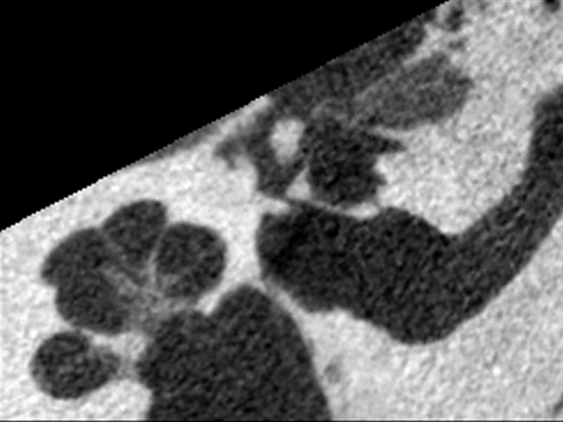
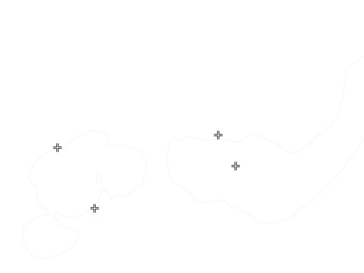 Hearing research · micro-CTKeypoints: landmarks an otologist placed, over the published outline.4 of 5 landmarks within 0.35 mm of this plane · round window (RW) is 2.5 mm off it, not drawnLabels on the picture: C · A · OW · V


Batteries, additive parts, food, rock cores, cultural heritage and more. Tell us what you inspect
Real scans and their labels, each modified (windowed, cropped, outlines drawn): Gruber et al., Fraunhofer EZRT, CC BY 4.0 (opens in a new tab); Kopp et al., MIT, CC0 (opens in a new tab); Armato et al., "Data From LIDC-IDRI", TCIA, CC BY 3.0 (opens in a new tab); van der Graaf et al., SPIDER, Radboud UMC, CC BY 4.0 (opens in a new tab); Amézquita et al., Michigan State University, CC0 (opens in a new tab); Wimmer et al., Bern University Hospital, CC BY 4.0 (opens in a new tab). Sources
Show us what you inspect.
Start with a public CT or MRI scan. See how a model suggestion becomes a reviewed label and a fixed training dataset.